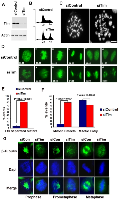
Figure 3. Timeless depletion causes mitotic chromosome defects.
A) HCT116 cells were transfected in duplicate with siControl or siTim and then assayed by Western blot at 24 hrs post transfection. Total cell extracts were probed with antibodies to Tim (top panel) or actin (lower panel). B) Cell cycle profiles of siControl or siTim transected HCT116 cells were generated by FACS analysis after propidium iodide staining. C) Metaphase spreads of siControl or siTim transfected HCT116 cells were generated at 24 hrs post-transfection. D) Images from time-lapsed micrographs of mitotic cell stages in siControl or siTim transfected HeLa cells that stabely express GFP-H2B to mark chromosomes. i) Prophase, ii) Prometaphase, iii) Metaphase, iv) Anaphase, v) Telophase, vi) Cytokinesis. E) Quantification of cells containing >10 separated sisters in metaphase spreads as represented in panel C (n = 86cells). Error bars represent standard deviation from the mean, and P values are derived from Chi-square analysis. F) Quantification of the number of mitotic defects (lagging chromosomes, failure to segregate, failure to progress to anaphase, failure during cytokinesis) observed for 5 independent movies (siControl, n = 207cells, siTim, n = 203 cells). Error bars represent standard deviation from the mean, and P values are derived from Chi-square analysis. G) Microtubule organization and centrosome disorganization in Tim depleted HeLa cells. Metaphase cells were stained for tubulin (green) by indirect immunofluorescence and DNA with Dapi (blue).

