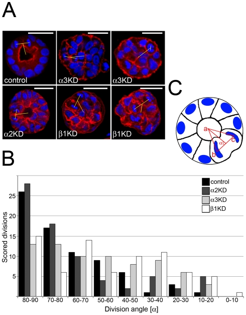
Figure 5. α3β1-integrin is required for mitotic spindle orientation.
A) Control, Itgα3-, Itgα2- and Itgβ1-KD MDCK cells were grown in 3D BME gel for 4 days, fixed and stained for nuclei (DAPI) and actin (TRITC-phalloidin). Confocal XY-sections from the middle of the cysts are shown. Sizebar is 30 µm. B) To analyze the orientation of cell division axis in cysts, stacks containing 74 mitotic post-metaphase cells from control, Itgα3-, Itgα2- and Itgβ1-KD cysts were collected. A frequency distribution of KD and control cells according to division angle is shown (P≤0.001 for Itgα3- and β1-KD). C) The XYZ-coordinates of cyst centre (a) and the segregated chromosomes of the dividing cell (b & c) were measured as shown in the schematic. The division angles (α) were calculated as described in materials and methods.

