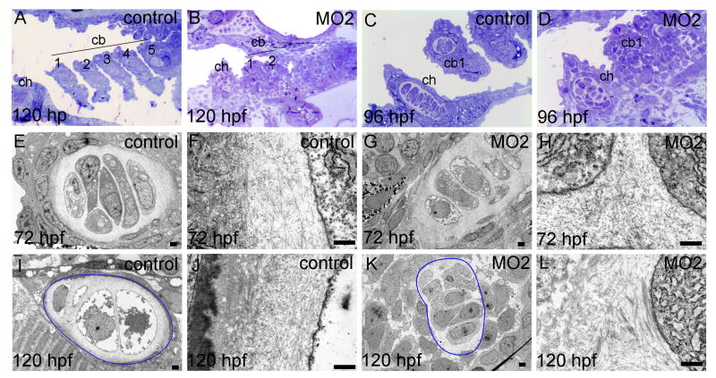
Fig. 5.
Chondrocyte morphology, stacking and ECM are abnormal in loxl3b morphants. Zebrafish embryos were injected with 2 ng controlMO or 2ng MO2. Transverse, semi thin sections were stained with Toluidine Blue for histological analysis (A-D). From the same embryos, ultrathin sections were analyzed using transmission electron microscopy to visualize chondrocyte morphology and ECM structure. In I and K, the outline of a ceratobranchial arch 1 is indicated in blue. ch: ceratohyoid, cb 1- 5: ceratobranchial arches 1 - 5. Scale bars indicate 500 nm (E-L).

