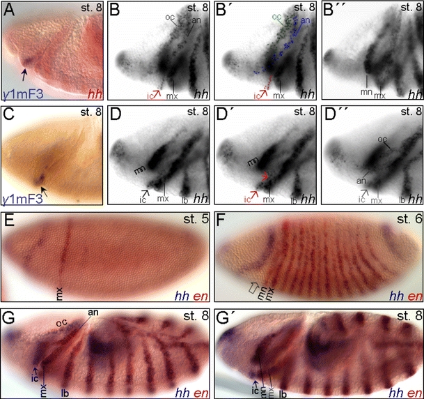
Fig. 6.

Early onset of the intercalary-specific expression of hh. A, C The γ1mF3 enhancer fragment (see Fig. 5A) mediates the early onset of reporter expression in the intercalary segment anlage (arrow) at stage 8 (see also Fig. 5M, N). B, B′′, D, D′′ Early procephalic expression of hh at stage 8. B, B′′ The procephalic stripes (oc, an, ic) are detected at a different focal plane (B, B′) than the mn stripe (B′′). B, B′′ In this lateral view, although detected at the same focal plane, the cell stripe in the intercalary (ic) segment anlage (arrow) is discontinuous from the antennal (an) segment anlage which progressively delineates from the ocular (oc) one. D, D′′ In this ventrolateral view, the cell stripes in the an and oc segment anlagen are detected at the same focal plane (D′′) while the ic is out of focus and vice versa. D, D' The slightly different focal plane of D′ compared to D allows the cell groups of both the ic anlagen to be detected. E–G′ Double in situ hybridization of hh (purple) and en (red). F Anterior to the mn stripe, the early procephalic expression domain of hh progressively splits into the antennal and ocular primordium during stage 7. The cells at the posterior margin of the early procephalic hh domain co-expressing en are precursors of cells of the antennal ectodermal stripe formed at the posterior procephalic margin at stage 8 (G). The open arrow depicts the precursor cells of the presumptive ic segment anlage. G, G′ Different focal planes of the same embryo at stage 8. hh but not en expression is detected in the ic segment anlage at stage 8 (arrow). For abbreviations, see Fig. 1
