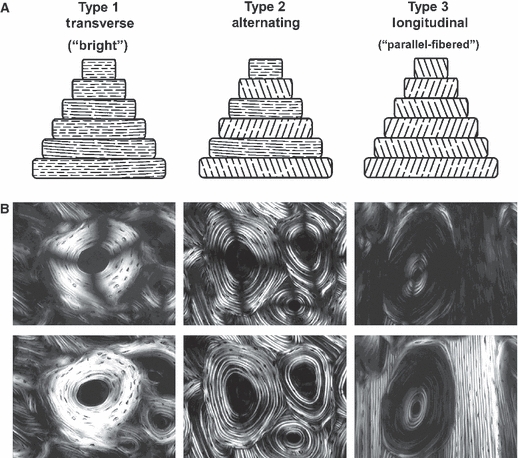
Fig. 2.

Examples of three-osteon morphotypes in linearly polarized light (top microscopic images) and circularly polarized light (bottom microscopic images; the dark cross-shaped extinction patterns are absent). The illustrations at the top of this figure are diagrammatic depictions of the predominant collagen fiber orientation (CFO) patterns that are believed be to the physical basis of the birefringence seen in the successive lamellae of the osteons in the microscopic images. The osteon images are from Bromage et al. (2003), and the layout of the figure is based on both Bromage et al. (2003) and Ascenzi & Bonucci (1968). These investigators ascribe to the view that the birefringence (gray-level) variations, seen for example in the ‘alternating’ osteon, are primarily based on lamellar variations in predominant CFO. Other investigators have argued that variations in the density of collagen fibers account for the gray-level variations in the alternating osteons (Marotti, 1996); this view is not well supported by experimental data and also appears to be influenced by limitations of specimen preparation techniques (Yamamoto et al. 2000). (Images are reproduced with permission of The Anatomical Record Part B, The New Anatomist; John Wiley and Sons Inc., Malden, MA, USA).
