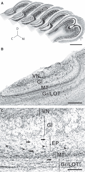
Fig. 1.

The accessory olfactory bulb of capybaras. (A) Sagittal representations throughout the olfactory bulb show that the accessory olfactory bulb (AOB) is located laterally. Note the size relation between the AOB and the main olfactory bulb (MOB). C, caudal; M, medial; D, dorsal. (B) Nissl-stained section revealing the cellular indentation between rostral and caudal subdivisions (dashed line) and AOB cell layers: VN, vomeronasal nerve; Gl, glomerular; MT, mitral/tufted cells; Gr/LOT, granule cells/lateral olfactory tract. (C) The extent of each layer is shown here with vertical lines. MT cells are densely packed at the MT layer and scatter in the inner portions of the external plexiform (EP) layer (arrowheads). Granule cells cluster at the Gr/LOT layer but also reach towards more superficial layers (arrows). Scale bars: 5 mm (A), 1 mm (B), 200 μm (C).
