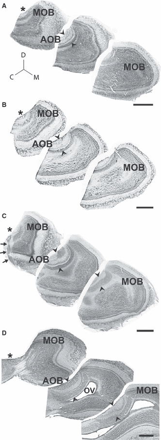
Fig. 4.

Lateral innervation and cell indentation in the AOB of caviomorph rodents. Panels A–D show Nissl-stained sagittal sections through the olfactory bulbs, from lateral portions on the left to progressively more medial portions on the right, of Ctenomys talarum (A), Spalacopus cyanus (B), Octodon lunatus (C) and Chinchilla laniger (D). The vomeronasal nerve arrives into AOB glomeruli from the lateral aspect (asterisks). Towards medial sections, all AOB glomeruli disappear (right). Note that a cell indentation is evident in all species (arrowheads). Nissl stain combined with immunohistochemistry for Gαi2-protein in O. lunatus shows the arrival of the vomeronasal nerve (arrows) and the coincidence of the cell indentation with rostral AOB expression of Gαi2-protein (C). Although the olfactory ventricle (OV) is collapsed in most caviomorphs, in chinchillas it is remarkably large. C, caudal; D, dorsal; M, medial; MOB, main olfactory bulb. Scale bar: 1 mm.
