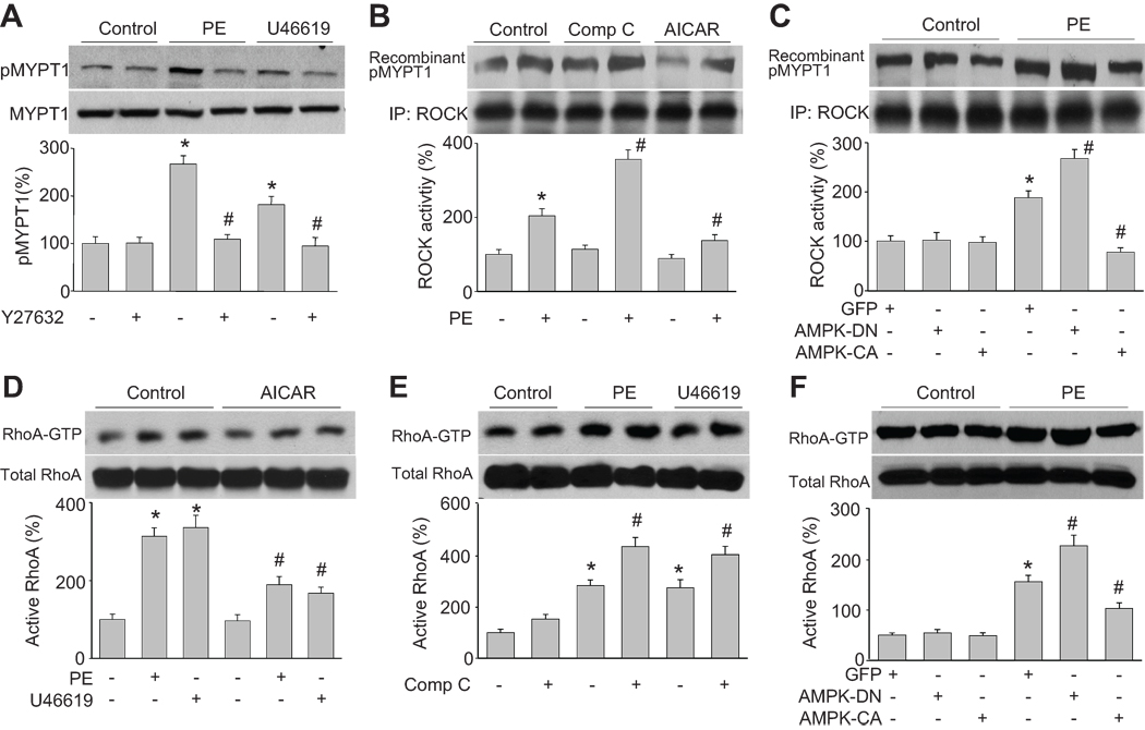
Figure 3. AMPK negatively regulates agonist-upregulated ROCK activity and RhoA activation.
(A) Western blot analysis of Thr696-phosphorylated MYPT1 in cultured HSMC pretreated with Y27632 (10 µM) for 2 h and then incubated with PE (1 µM) or U46619 (30 nM) for 30 min. n=3, *P<0.05 vs. control, #P<0.05 vs. PE or U46619 alone. (B) ROCK activity in cells were treated with AICAR (2 mM) or compound C (20 µM) for 2 h and then incubated with PE for 30 min. Levels of Thr696-phosphorylated recombinant MYPT1 served as an index of ROCK activity. n=3, *P<0.05 vs. control, #P<0.05 vs. PE alone. (C) ROCK activity in cells transduced with adenovirus vectors encoding GFP, AMPK-DN (DN), or AMPK-CA (CA) for 48 h and then treated with PE (1 µM) for 30 min. n=3, *P<0.05 vs. GFP alone, #P<0.05 vs. GFP plus PE. (D) RhoA activation, as measured by Rhotekin pull-down, in cells were pretreated with AICAR (2 mM) for 2 h and then incubated with PE (1 µM) or U46619 (30 nM) for 30 min. n=3, *P<0.05 vs. control, #P<0.05 vs. PE or U46619 alone. (E) RhoA activation in cells pretreated with compound C (20 µM) for 2 h and then incubated with PE (1 µM) or U46619 (30 nM) for 30 min. n=3, *P<0.05 vs. control, #P<0.05 vs. PE or U46619 alone. (F) RhoA activation in cells transduced with endovirus vectors encoding GFP, AMPK-DN (DN), or AMPK-CA (CA) for 48 h and then treated with PE (1 µM) for 30 min. n=3, *P<0.05 vs. GFP alone, #P<0.05 vs. GFP plus PE.

