
Figure 2.
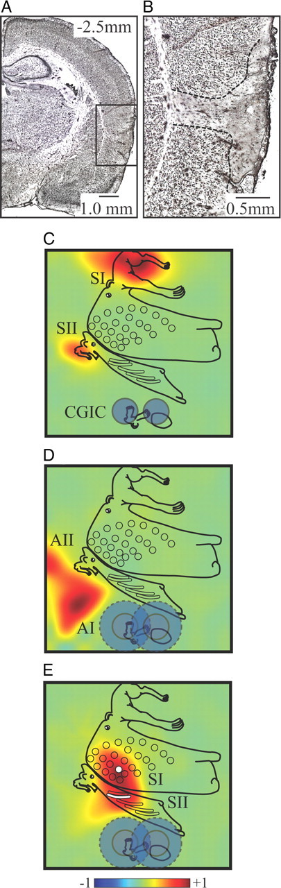
Anatomical and functional verification of excitotoxic CGIC lesions. A, Photomicrograph showing the extent of the excitotoxic lesions in an example at the most caudal site. Scale bar, 1.0 mm, NeuN stain. B, Enlargement of the lesion site shows cell death restricted to the cortical mantle and not crossing the external capsule into the underlying striatum. Scale bar, 0.5 mm. C, Functional verification of lesions (dark blue circles denote anatomical lesion extent) indicates complete suppression of the SEP in CGIC but leaves responses in both SI and SII unaltered. D, AEPs are completely eliminated in auditory CGIC and attenuated in the most rostral region of AI but are unaffected in AII, suggesting an area of functional lesion closer to 2 mm in diameter (light blue circles). E, Functional verification of SEPs evoked by stimulation of B2 whisker (the B2 representation in SI is indicated by white circle and in SII by a white bar) shows the intact whisker response in SII, just dorsal to the lesion site, demonstrating the confined distribution of the lesion.