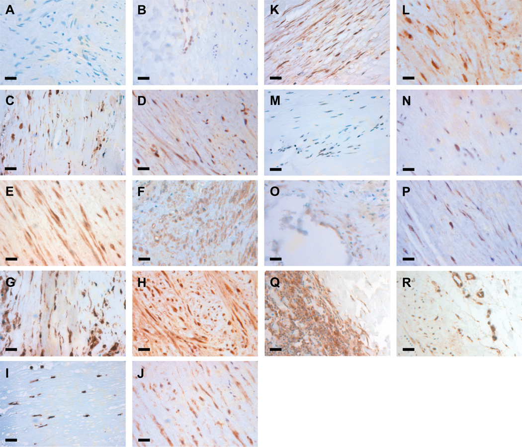
Figure 4.
Immunohistochemical staining for transcription factors with enriched binding sites. Immunostaining was performed in both AAA tissue (first and third column) and non-aneurysmal control aorta (second and fourth column) for PEA3 (A,B), ELF1 (C,D), ETS2 (E,F), ETS1 (G,H), NFKB p65 (I,J), NFKB p50 (K,L), GABP alpha (M,N), AML1 (O,P), and STAT1 (Q,R). Positive staining is seen for all proteins in both AAA tissue and non-aneurysmal control aorta with the exception of PEA3. Bars=20 µm.

