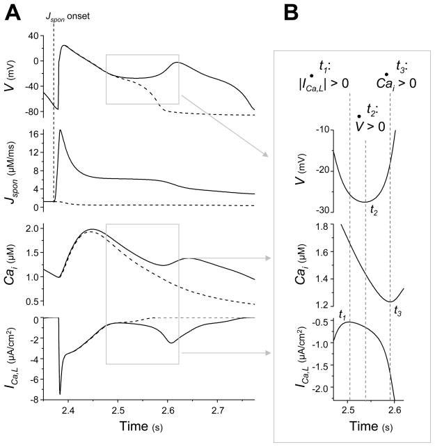
Fig. 2.
A low amplitude, long duration Jspon event starting during late diastole causes an ICa,L-induced (“early”) EAD. (A) Traces of V, Jspon, Cai, and ICa,L in time during a tachycardia-induced EAD in an AP model with the low amplitude, long duration Jspon (solid lines). After stimulating the AP model at a rate of 160 ms for approximately 2.4 s, a large Jspon activation starts in late diastole 8 ms before the next paced AP (vertical dotted line labeled “Jspon onset” indicates start of Jspon). An EAD occurs during the repolarization phase of the ensuing AP (t ≈ 2.6 s), approximately 170 ms after the Jspon onset. A Cai aftertransient and ICa,L reactivation are observed in conjunction with the EAD. Jspon is small but nonzero during the EAD itself. Dashed traces show the result of preventing the large Jspon activation by setting p∞ = 0 just before it would otherwise occur. (B) Enlargement of the traces in (A) during EAD upstroke, illuminating the exact sequence of events. Time derivatives are denoted with dots (i.e., dV/dt = V̇). Slow ICa,L reactivation (indicated by d(|ICa,L|)/dt > 0) occurs first, at time t1. The window current increases inward current and reverses repolarization, inducing the V upstroke beginning at time t2 (indicated by dV/dt > 0). The increasing V has a positive feedback effect on ICa,L, causing rapid reactivation and driving a Cai upstroke (indicated by d(Cai)/dt > 0) at time t3.

