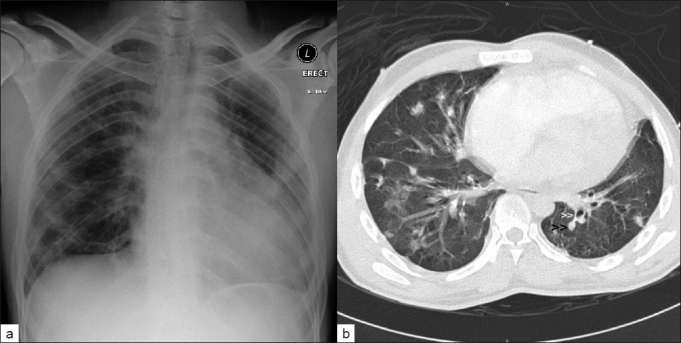
Figure 1:

(a) Chest X-ray showing cardiomegaly with bilateral lung infiltrates and (b) CT scan of chest showing bilateral irregular pulmonary infiltrates with multiple nodules (black arrowheads) with distinct central feeding vessel (white arrowheads), consistent with septic pulmonary emboli in a patient with intravenous drug abuse and tricuspid valve endocarditis
