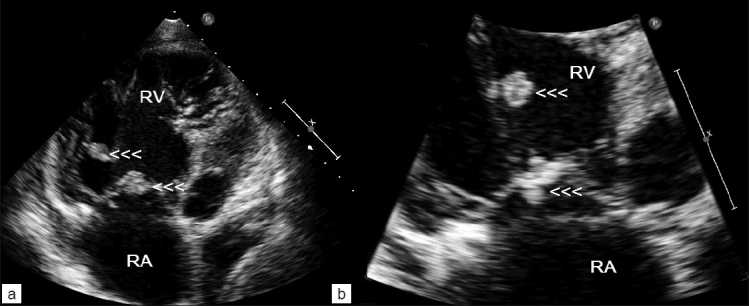
Figure 2:

Transthoracic echocardiography (a and b) showing multiple vegetations attached to tricuspid valve leaflets (arrowheads) and one large vegetation on the chordae of anterior tricuspid leaflet (upper arrowheads) in a patient with IV drug abuse and septic pulmonary emboli. RA, right atrium; RV, right ventricle
