
Figure 1.
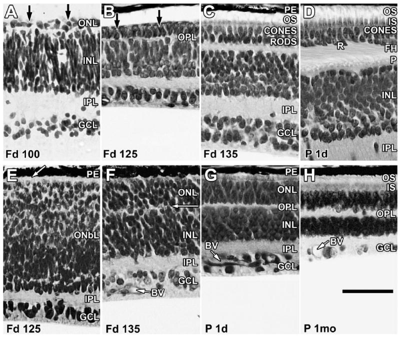
Layer names and their abbreviations given in Figure 1 are used throughout the figure legends. Sections of marmoset retina at the foveal edge (A–D) and in the nasal midperiphery (E–H) stained with azure II and methylene blue. A: At Fd 100, the retina contains a distinct outer nuclear layer (ONL), a thin outer plexiform layer (OPL), a thick inner nuclear layer (INL), a thick inner plexiform layer (IPL), and a ganglion cell layer (GCL). The ONL contains cones and rods (arrows) but is still very thin. B: At Fd 125, the foveal edge has distinct cones and rods (arrows), and cell types are identifiable in INL. C: By Fd 135, the ONL contains more rods and is thicker because of the onset of cone and rod packing. Short outer segments (OS) are present. D: At birth, both rods (R) and cones have formed basal synaptic axons or FH, which tilt away from the foveal center (to the left), marking laminar displacement during pit formation. Both OS and inner segments (IS) are longer. The OPL is thicker, with a solid row of cone pedicles (P) lining its inner edge. Cones now are two or three deep, with rods intermixed between the cone FH. E: At Fd 125, the peripheral retina is markedly less mature than the central retina. The inner retina has a distinct IPL and GCL, but the outer retina is a thick neuroblastic layer (OnbL) in which only cones can be distinguished at the outer edge (arrow). F: By Fd 135, the ONbL is subdivided into ONL and INL by a thin, irregular OPL (arrow). Rods and cones are still difficult to identify in the ONL. Blood vessels (BV) in the GCL are growing across the peripheral retina. G: All layers are present in peripheral retina at birth, and BV have reached far peripheral retina. The ONL contains an outer layer of cones and a thick inner layer of rods that have short IS and OS in the interphotoreceptor space between the ONL and the pigment epithelium (PE). H: By 1 month, peripheral IS and OS are much longer, and the retina appears mature. Scale bar = 50 μm.