
Figure 4.
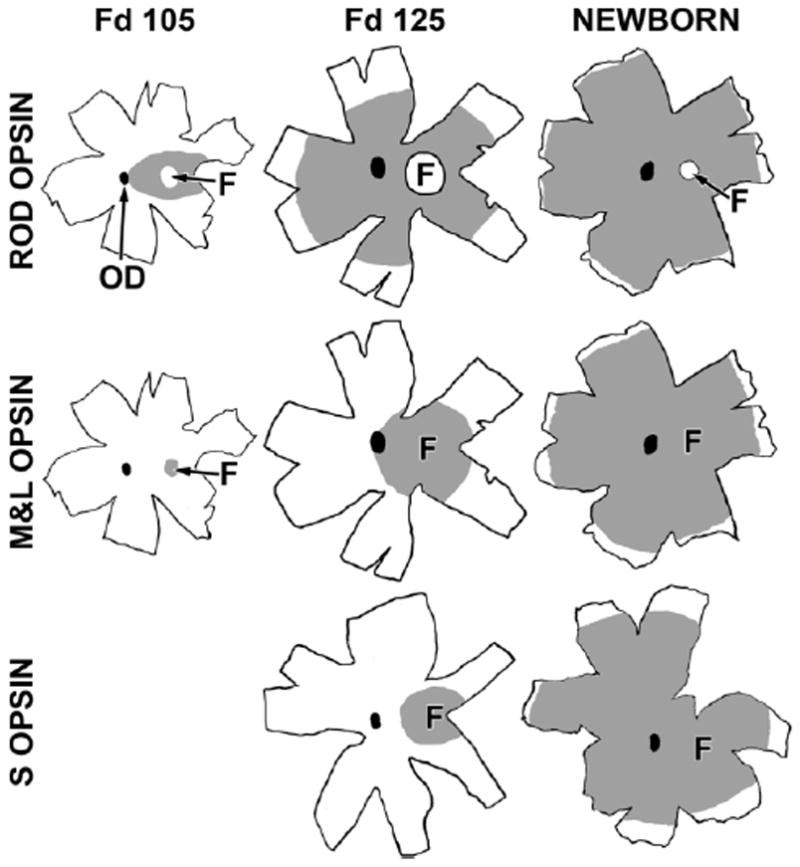
Drawings of marmoset retinal whole mounts immunocytochemically labeled for rod opsin (top row), M&L cone opsin (middle row), and short-wavelength-selective (S) cone opsin (bottom row). The optic disc (OD) is the black oval in each drawing. The gray shading is the region containing labeled photoreceptors at each age. Rod opsin covers the central retina at Fd 105, with the exception of the fovea (F, indicated by the arrow). By Fd 125, rod opsin covers 70% of the retina, and, at birth, many rods near the retinal edge are labeled. Note that the fovea remains rod-free throughout development and that it becomes smaller with age. M&L opsin at Fd 105 is present only in the foveal cones. By Fd 125, M&L+ cones fill the central retina to the OD, and, at birth, M&L opsin is near, but not at, the retinal edge. In contrast, at Fd 105, very few S opsin+ cones were detected within the fovea (not shown). By Fd 125, S opsin was present only in and around the fovea and was still relatively far from the edge at birth. Scale bar = 5 mm.