
Figure 5.
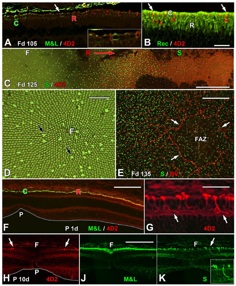
Immunocytochemical labeling showing the expression of cone and rod opsins. A: The edge of the Fd 105 incipient fovea, showing the first M&L+ cones (C) and rhodopsin+ (4D2) rods (R). Note that both types of opsin+ photoreceptors are scattered and not adjacent to one another. The region indicated by the arrow is shown at higher magnification in the inset. A small 4D2+ red rod is adjacent to a larger thicker M&L+ green cone. Note that opsin labeling is mainly in the cell membrane and tiny OS. B: The edge of 4D2 expression near the optic disc at Fd 105. Many rods (R) are Rec+, whereas only a few have 4D2 (arrows). C: A Fd 125 whole mount comparing S opsin and 4D2 expression. The fovea (F, left) contains an irregular mosaic of S+ cones that extends for >1.5 mm outside the fovea (S). The first rods (R) appear on the foveal edge. Many 4D2+ rods are present into the periphery (direction of arrow), and 4D2+ rods extend for at least 3 mm to the right of the picture edge. This pattern demonstrates that rod opsin is expressed in advance of S opsin. D: Higher magnification view of the fovea in C. The foveal cone mosaic radiates from the foveal center (F). S+ cones (black arrows) are irregularly scattered throughout the more numerous smaller M&L cones. E: Blood vessels (BV) forming the foveal avascular zone (FAZ; arrows) outline the incipient fovea. S+ cones are present at low density within the FAZ and at higher density outside it. F: At birth, the immature foveal pit (P) is shallow and wide (dotted line). Overlying the pit, M&L+ cones (C) form a single layer that extends for 400–450 μm to where the first 4D2+ rods (R) appear. G: The 4D2+ rods on the neonatal foveal edge have short FH (arrows) that point away from the foveal center, marking bipolar cell displacements in the INL associated with pit formation. H,J: Serial sections through the P10d fovea showing that the pit now is broader (H, dotted line) and the most central 4D2+ rods (arrows) are >200 μm from the foveal center (F). M&L+ cones are packed into the foveal center, forming a central multilayered ONL (J). S+ cones extend across the foveal center (K). These also have FH that point away from the foveal center (inset). A magenta-green copy is available as Supporting Information Figure 1. Scale bars = 50 μm in B (applies to A,B); 500 μm in C; 100 μm in D,E; 200 μm in F; 50 μm in G; 100 μm in J (applies to H–K).