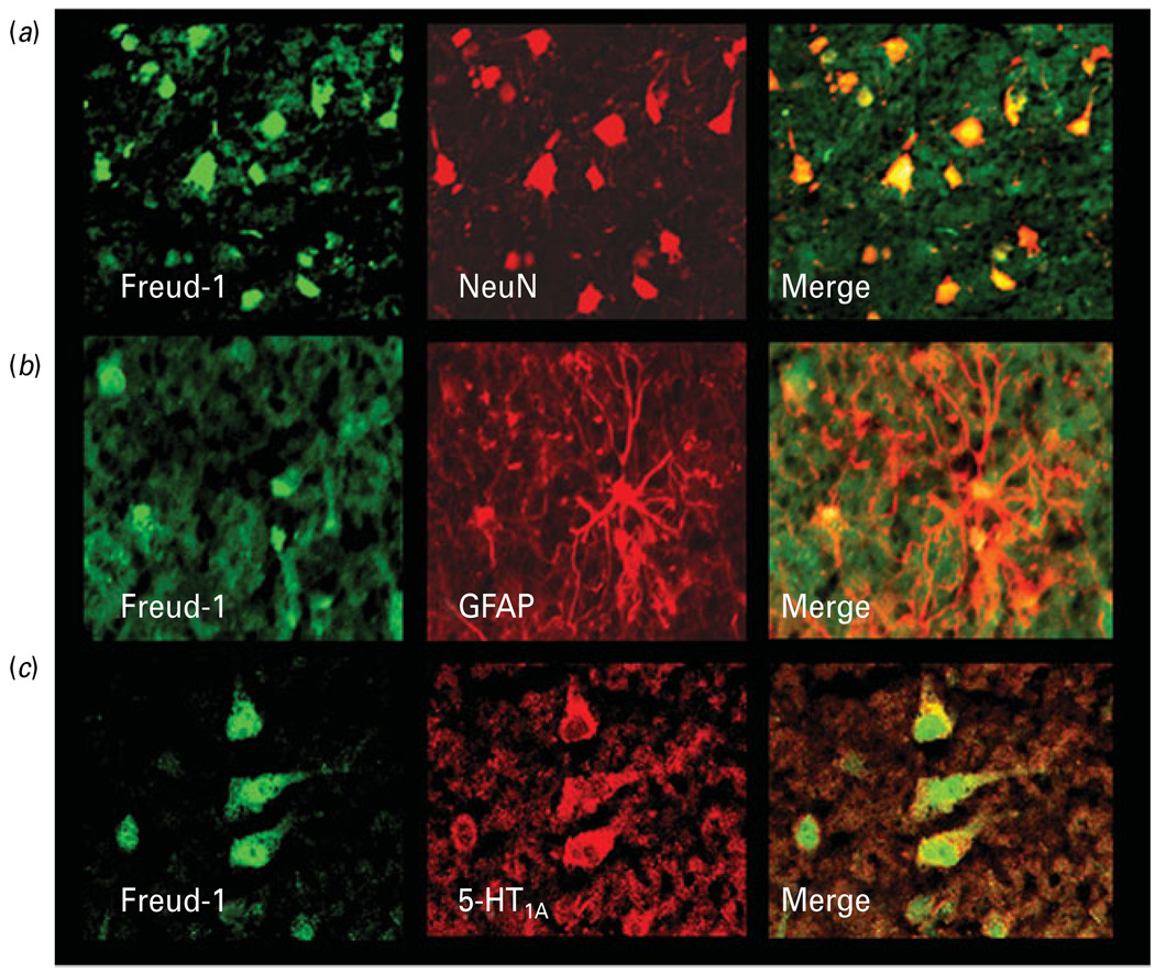
Fig. 3.
Colocalization of Freud-1 immunoreactivity (IR) in neurons and glia. (a) Co-labelling of Freud-1 IR (green) with the neuronal marker NeuN (red). Freud-1 protein (yellow) is localized to neuronal nuclei and perinuclear areas. (b) Co-labelling of Freud-1 IR (green) with the astrocytic marker GFAP (red). Freud-1 (yellow) is localized in glial cell nuclei. (c) Colocalization of Freud-1 IR (green) and immunoreactivity for 5-HT1A receptor (red). Freud-1 IR (yellow) is present in the cytoplasm of a majority of cells expressing 5-HT1A receptor immunoreactivity.

