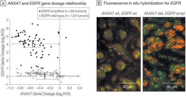
Figure 1. Relationship Between the ANXA7 and EGFR Genes (193 Tumors).
A, Gene dosage relationship for annexin A7 (ANXA7) and epidermal growth factor receptor (EGFR) in glioblastomas from The Cancer Genome Atlas (TCGA) pilot project. Analysis included 193 of 219 TCGA samples for which all EGFR probes on the Agilent aCGH array yielded consistent amplification or wild-type (wt) calls. Scatterplot shows the relationship between EGFR and ANXA7 gene dosage along the Cartesian coordinates for each of the 193 tumors. Gene dosage values indicate the log2 ratio of red-to-green fluorescence dye intensity (log2R/G) estimated by the circular binary segmentation algorithm. Negative gene dosage indicates lower gene dosage compared with that observed for the majority of the samples, ie, lower gene dosage compared with a normal diploid state. Boxplots depict the first quartile minus 1.5 × interquartile range (IQR) (left whiskers) and the third quartile plus 1.5 × IQR (right whiskers), IQR (box), and median (vertical line); 1 observation outside the whiskers indicates an outlier. Wilcoxon rank-sum testing for the difference in ANXA7 gene dosage in EGFR-amplified (ampl) vs wild-type tumors discloses a significantly diminished ANXA7 gene dosage in the EGFR-amplified tumors (P=.000000001). B, Representative fluorescence in situ hybridization analysis of EGFR gene dosage in 1 ANXA7–wild-type and 1 ANXA7-deleted (del) glioblastoma, from the Stanford set of tumors, on paraffin. Red dots (Spectrum Orange dye) are specific for the EGFR gene locus probe and green dots (Spectrum Green dye) arise from a probe specific for the centromeric region of chromosome 7. The ANXA7–wild-type tumor is nonamplified for EGFR gene dosage with most cells showing normal diploid levels of 2 red and 2 green signals per cell, while the ANXA7-deleted tumor is amplified for EGFR gene dosage as the cells show a robust increase in the number of EGFR red dots. Additionally, the ANXA7-deleted tumor is polyploidic for chromosome 7 as there are more than 2 green dots (centromere of chromosome 7) in most cells (original magnification ×40).

