Figure 1.
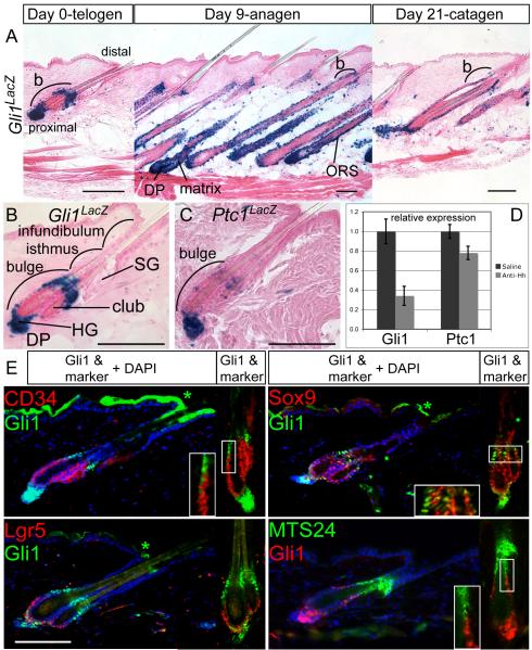
Hh-responding cells are localized in molecularly distinct subdomains of the telogen hair follicle. Also see Figures S1, S2.
(A) X-gal staining in adult Gli1LacZ/+ skin at day 0, 9, and 21 after depilation of telogen hair to induce regeneration of the anagen follicle. b; bulge, Scale bars=100μm.
(B, C) X-gal staining of Gli1LacZ/+ and Ptc1LacZ/+ telogen follicles showing Hh-response genes in the upper bulge, lower bulge, HG, and DP. Scale bars=100μm.
(D) Relative expression levels of Gli1 and Ptc1 mRNA assessed by RT-qPCR in wild type telogen skin treated with Hh-neutralizing antibody. Error bars=SEM. club; club hair.
(E) Immunostaining of Gli1LacZ and the progenitor cell markers indicated. Blue; DAPI. *Non-specific staining. Scale bar=100μm.

