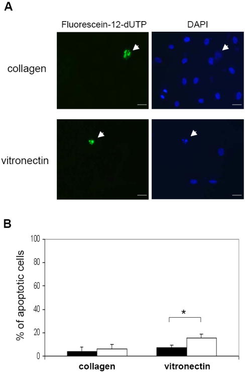
Figure 3. HKa fails to induce apoptosis of EPCs on collagen-coated surface.

EPCs were cultured on plates precoated with collagen or vitronectin in the presence or absence of 50 nM HKa. After 72 hours, cell apoptosis was analyzed with the TUNEL method as described in the Materials and Methods. (A) Representative fluorescent micrographs of Fluorescein-12-dUTP-positive cells (left) and counterstaining with DAPI (right) in HKa-treated EPCs. Scale bar represents 25 μm. (B) EPCs were cultured with 0.1% BSA or 50 nM HKa on collagen or vitronectin surfaces for 72 hours, which was followed by TUNEL assay. Percentage of apoptotic cells is shown. Closed column, BSA; Open column, HKa. *P<0.01 v.s. BSA.
