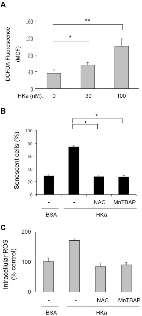
Figure 5. HKa stimulates generation of ROS in EPCs.

(A) H2DCF-DA oxidation in EPCs. As described under Materials and Methods, EPCs were preincubated with 10 μM H2DCF-DA for 30 minutes, and then treated with or without HKa for 12 hours. H2DCF-DA oxidation was analyzed by flow cytometry. The data are expressed as the mean channel fluorescence (MCF) of H2DCF-DA and represent the average of three separate experiments (± SEM).*P<0.05, **P < 0.001. (B and C) Effect of NAC and MnTBAP on HKa-induced EPC senescence (B) and ROS production (C). EPCs were cultured in the absence or presence of 50 nM HKa for 14 days. As indicated, EPCs in separate groups were also treated with 0.1% BSA (-), 100 μM NAC or 10 μM MnTBAP. Percentage of senescent cells was calculated and depicted (B), and intracellular ROS level are expressed as percent of controls (*P < 0.001, v.s. BSA; n=3).
