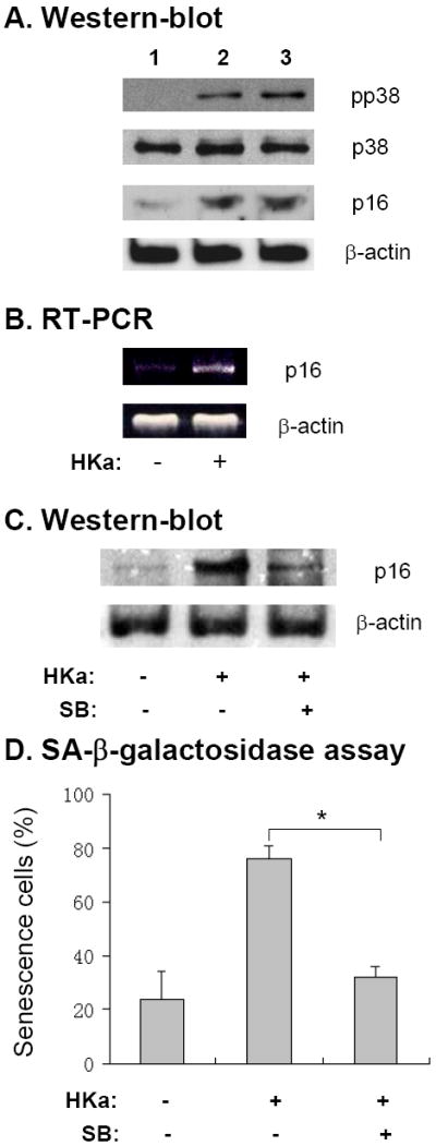
Figure 6. HKa increases p38 kinase activity and p16INK4a expression in EPCs.

(A) EPCs were cultured in the presence of 0.1% BSA (lane 1), 30 nM HKa (lane 2) or 100 nM HKa (lane 3) for 14 days. Representative immunoblot of phosphorylated p38 kinase and p16INK4a (p16) level in EPCs is shown. The blots for p38 kinase and β-actin serve as loading control. (B) RT-PCR analysis of p16INK4a (p16) mRNA expression in EPCs, which were cultured with or without 50 nM HKa for 14 days. Representative agarose gel image from three experiments is shown. (C) EPCs were cultured in the presence or absence of 50 nM HKa with or without 10 μM SB203580 for 14 days. The level of p16 expression was analyzed by immunoblotting. Representative result from three experiments is shown. The blot for β-actin serves as loading control. (D) EPCs were cultured as described in the panel C. Percentage of senescent EPCs was calculated by SA-β-galactosidase assay (* p<0.01; n=3).
