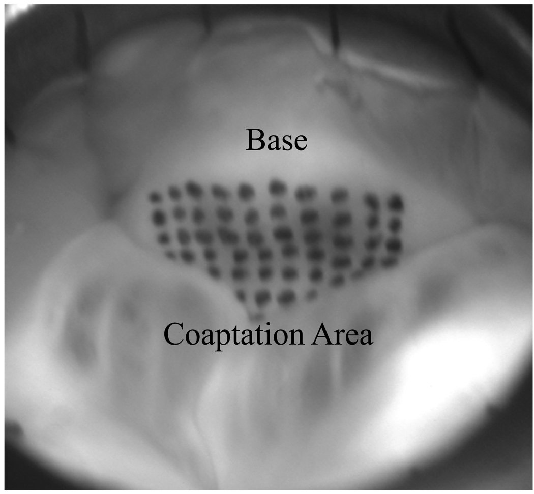
Figure 2. Ventricular View of Leaflet with Marker Grid.
The whole aorta was sutured into the flow loop such that the ventricular sides of the leaflets were visible to be photographed. The ventricular side of the non-coronary leaflet was marked with a grid of 48 markers using tissue marking dye but the coaptation geometry allowed only 42 visible markers.

