
Figure 4.
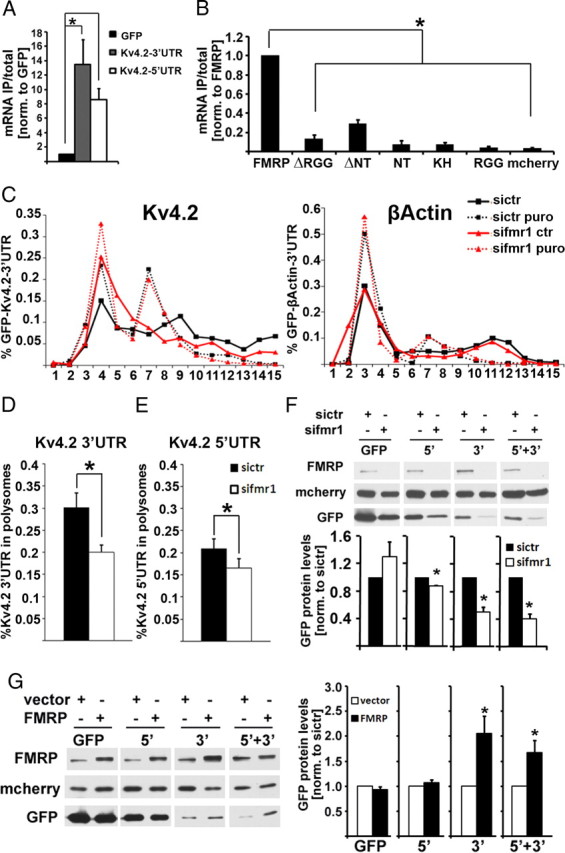
FMRP regulation of Kv4.2 mRNA is mediated by both 5′- and 3′-UTRs of Kv4.2 mRNA. A, The association of both GFP-Kv4.2 3′-UTR and Kv4.2 5′-UTR-GFP mRNAs with mcherry-FMRP protein is significantly higher than that with GFP mRNA alone (n = 4, one-way ANOVA with least significant difference post hoc tests: *pGFP 3′-UTR = 0.007, *pGFP 5′-UTR = 0.049). B, mcherry-FMRP shows significantly higher affinity to the 3′-UTR of Kv4.2 mRNA than does mcherry-FMRP lacking either the RGG box (ΔRGG) or the N terminus (ΔNT), or mcherry-fusion proteins containing either of the three known FMRP RNA-binding domains [N terminus (NT), KH-domain, or RGG-box, respectively] (n = 6, one-way ANOVA, *p < 0.0001; Tukey's honestly significant difference post hoc comparisons, *p ≤ 0.05). C, Association of recombinant GFP-Kv4.2 3′-UTR (16 h expression) with puromycin-sensitive polysome fractions is reduced in HEK293T cells after 48 h of siRNA-mediated depletion of FMRP (left), whereas the β-actin 3′-UTR is unchanged (right). D, Quantitative analysis shows a significant reduction of Kv4.2 3′-UTR in heavy polysomes from FMRP-knockdown cells (n = 4, *p = 0.0195, paired t test). Similar results were seen for a luciferase reporter, and in N2A cells (data not shown). E, Likewise, Kv4.2 5′-UTR was reduced in polysomes from FMRP-depleted HEK293T cells, but to a lesser extent compared with the 3′-UTR (n = 3, *p = 0.031, paired t test; 3′-UTR, 33%; 5′-UTR, 21% reduction). F, GFP protein levels are reduced when fused to either Kv4.2 5′-UTR (n = 3, *p = 0.003, paired t test), Kv4.2 3′-UTR (n = 3, *p = 0.021, paired t test), or both UTRs (n = 3, *p = 0.017, paired t test) in FMRP-knockdown cells compared with control knockdown cells; however, a GFP construct without any UTRs (n = 3, p = 0.297, paired t test) shows no significant expression difference (72 h of siRNA treatment, 16 h coexpression of GFP-constructs with mcherry). Representative Western blot images are shown above; mcherry was used as transfection control. G, Overexpression of FMRP leads to a significant increase of GFP protein levels fused to both UTRs or the Kv4.2 3′-UTR (n = 7; GFP, p = 0.301; 5′, p = 0.303; 3′, *p = 0.023; 5′ + 3′, *p = 0 .033, paired t test). Representative Western blots are shown on the left. All error bars represent SEM.