
Figure 5.
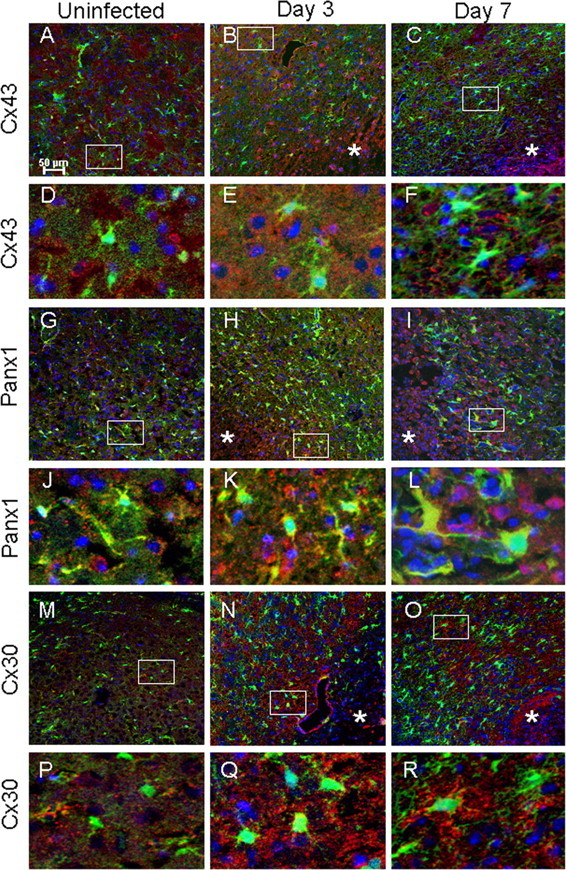
Inflammation alters Cx43, Panx1, and Cx30 expression in brain abscesses. Tissues from uninfected (A, D, G, J, M, P) GFAP–GFP mice or animals infected with S. aureus at either day 3 (B, E, H, K, N, Q) or 7 (C, F, I, L, O, R) after infection were stained for Cx43, Panx1, or Cx30 (red) and 4′,6′-diamidino-2-phenylindole (DAPI) (blue), whereas GFAP–GFP+ astrocytes are green and analyzed by confocal microscopy. Brain abscess margins are denoted with asterisks, and representative staining from the contralateral uninfected hemisphere is presented to assess basal expression in the striatum. The insets in A–C represent areas of higher magnification that are presented in D–F, respectively, to demonstrate Cx43 cellular localization. Similarly, the insets in G–I represent areas that are shown in J–L, respectively, to depict Panx1 cellular localization, and the insets in M–O correspond to P–R to represent Cx30 cellular localization.