
Figure 6.
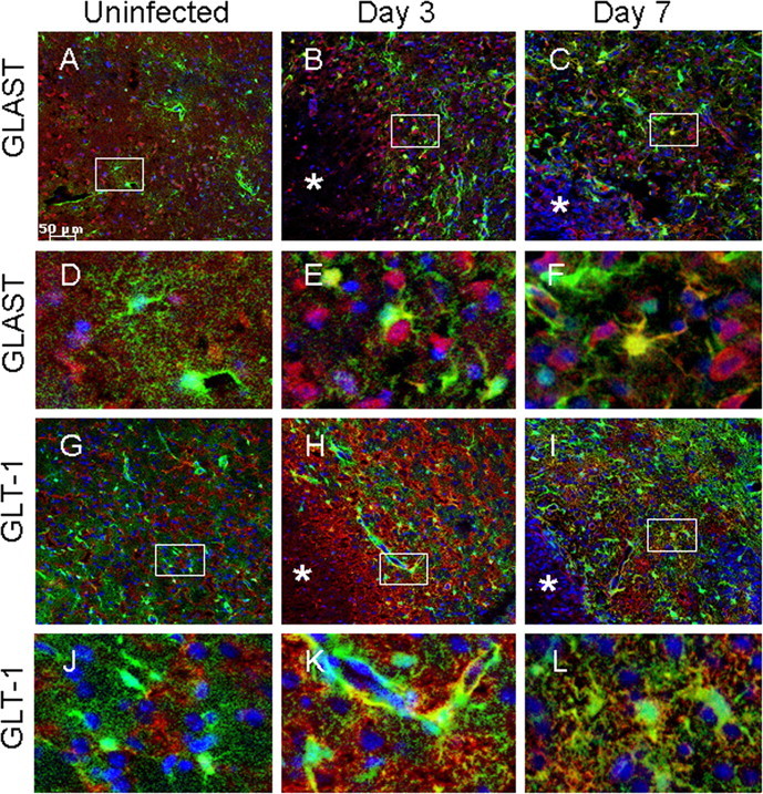
Expression of the glutamate transporters GLT-1 and GLAST is altered in response to the inflammatory milieu. Tissues from uninfected (A, D, G, J) GFAP–GFP mice or animals infected with S. aureus at either day 3 (B, E, H, K) or 7 (C, F, I, L) after infection were stained for GLT-1 or GLAST (red) and DAPI (blue), whereas GFAP–GFP+ astrocytes are green and analyzed by confocal microscopy. Brain abscess margins are denoted with asterisks, and representative staining from the contralateral uninfected hemisphere is presented to assess basal glutamate transporter expression in the striatum. The insets in A–C represent areas of higher magnification that are presented in D–F, respectively, to demonstrate GLAST cellular localization. The insets in G–I represent areas of higher magnification that are presented in J–L, respectively, to depict GLT-1 cellular localization.