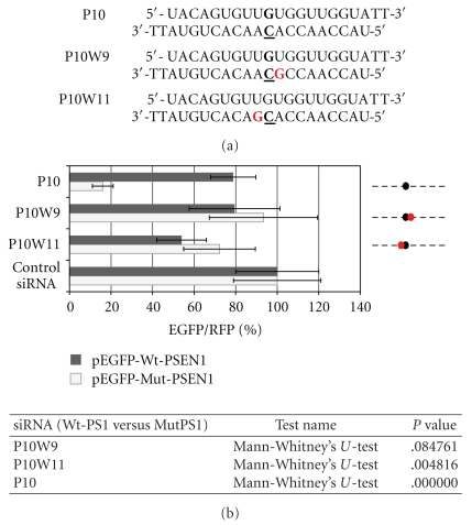
Figure 9.
(a) Sequences of the P10W9 and P10W11 siRNAs modified in the central domain at the position 9 or 11; A is replaced by G (in red) to generate the wobble base pair with complementary mRNA strand. (b) Comparison of the florescence levels obtained after co-transfection of HeLa cells with pEGFP-Wt-PSEN1(400) or pEGFP-Mut-PSEN1(400), pDsRed-N1 and indicated siRNAs (P10, P10W9, or P10W11). Dotted lines represent the antisense strands of the used siRNAs. Positions of the C-C mismatch between the guide strand of siRNA and the wild-type PSEN1 mRNA are indicated by the black dots. Red dots represent the sites of the wobble base pairs between the guide strand of siRNA and the wild- or mutated-type of PSEN1 mRNA. The level of the relative EGFP/RFP fluorescence in cells transfected with control nonsilencing siRNA was used as 100%. The results are mean values from three independent experiments. Statistical analysis of differences between two groups of data (Wt-PSEN1 versus Mut-PSEN1) were calculated by the use by the nonparametric Mann-Whitney's U-test. The differences with P < .05 were considered as statistically significant.

