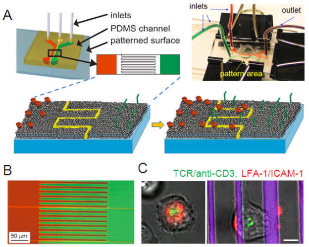
Figure 4. Multicomponent supported lipid bilayers.
(A) Microfluidic approaches allow patterning of supported lipid bilayers at subcellular levels. (B) Example of a two-component lipid bilayer system. (C) Comparison of T cell receptor organization on unpatterned (left) and segregated (right) lipid bilayers presenting ligands to TCR and LFA-1; scale bar = 5 μm. Adapted from [58].

