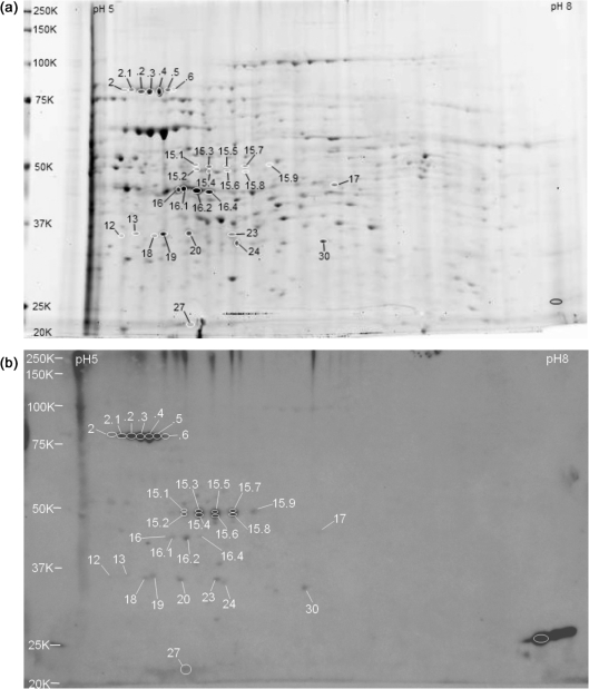
Fig. 4.
Whole-cell extracts of C. burnetii RSA 493 phase I grown in mouse L929 cell culture and separated by 2D gel electrophoresis on pH 5–8 IEF gradients and 10.5–14 % acrylamide SDS-PAGE gels. C. burnetii seroreactive proteins are listed in Table 4. Molecular mass standards (kDa) are indicated on the left. (a) Sypro Ruby-stained gel showing seroreactive spots. (b) Immunoblot probed with pooled immune guinea pig serum.

