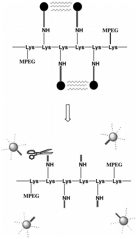
Fig. 2.
Schematic shows design of the MMP-2-sensitive fluorescent probe for in vivo NIRF imaging. Fluorophores with excitation and emission wavelengths in the NIR spectrum were covalently coupled to a poly-l-lysine backbone (—Lys-Lys-Lys…—) sterically protected by methoxy polyethylene glycol (MPEG) side chains by means of a synthetic MMP-2 peptide substrate. Top: Owing to the proximity of the fluorophores, fluorescence resonance energy transfer occurs so that almost no fluorescent signal can be detected in the nonactivated state. Bottom: After MMP-2 cleavage of the peptide spacer, fluorophores are released from the carrier and become brightly fluorescent. Figure is reproduced and modified with permission from Radiology [53]. “Copyright (2001) The Radiological Society of North America (RSNA)”.

