Figure 2.
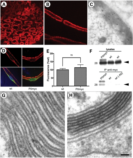
P0ct-myc recapitulates P0wt function. (A and B) IHC for myc (red) in transverse sections (A) and teased fibres (B) from P0ct-myc sciatic nerves shows that P0ct-myc is located in myelin. (C) IEM analysis for myc in P0ct-myc sciatic nerves reveals that P0ct-myc is distributed throughout the myelin sheath. (D and E) Confocal microscopic analysis of teased fibres reveals similar intensity of P0 signal (red) in perinuclear KDEL-positive (green) Schwann cell cytoplasm in P0ct-myc when compared with wt sciatic nerves (note that the P0 antibodies recognize both P0wt and P0ct-myc as in Figure 1C; see Supplementary Material, Figure S2, for sampling area, and Figure S3 for S63del-positive control); ns, not significantly different. (F) Anti-myc antibodies co-immunoprecipitate P0wt (Mr = 28, arrowhead) from P0ct-myc sciatic nerve lysates. wt lysates in IP with myc antibody or with only beads [no myc antibody, wt (−Ab)] serve as negative controls. (G and H) Ultrastructural analysis of myelin sheaths from P0ct-myc/Mpz−/− nerves (no P0wt present) shows regular presence of several compacted myelin wraps (H), which are found much less often in Mpz−/− nerves (G) (Table 1). P0myc, P0ct-myc. Scale bar in H (A and B): 9 μm; (C): 200 nm; (D): 26 μm; (G and H) 50 nm.

