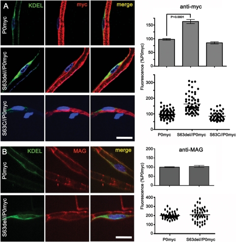
Figure 3.
P0S63del induces ER retention of P0ct-myc, but not MAG. (A and B) Teased fibres from P0ct-myc, S63del//P0ct-myc or S63C//P0ct-myc sciatic nerves were stained for DAPI (blue), KDEL (green) and myc (A; red) or MAG (B; red) and imaged by confocal microscopy. The intensity of myc or MAG staining was measured in comparable perinuclear KDEL-positive areas and expressed as the percent of intensity for P0ct-myc. P0myc, P0ct-myc. Scale bars: 20 μm.

