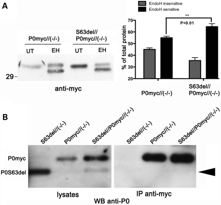
Figure 4.
In the presence of P0S63del, P0ct-myc shows more glycosylation typical of the ER, but does not co-immunoprecipitate with P0S63del. (A) Western analysis for myc was performed on EndoH-treated (EH) or -untreated (UT) sciatic nerve lysates of P0ct-myc//Mpz−/− or S63del//P0ct-myc//Mpz−/− mice. EndoH-sensitive and -insensitive bands from four sciatic nerve lysates (four mice) of each genotype were quantified by densitometry and expressed as %total signal. (B) IP of myc followed by western analysis for P0 on SN lysates does not detect P0S63del (arrowhead), whereas direct western analysis for P0 does. P0myc, P0ct-myc.

