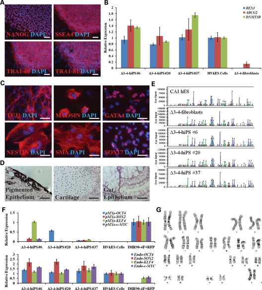
Figure 2.
Δ3–4-hiPS cells are pluripotent and fully reprogrammed. (A) Δ3–4-hiPS #37 express pluripotency markers by immunocytochemistry. Scale bars, 100 µm. (B) Δ3–4-hiPS cells express bona fide hiPS cell markers by qRT–PCR. Data are expressed as mean ± SEM. (C) Δ3–4-hiPS #37 differentiate into the three germ layers, ectoderm (TUJ1, NESTIN), mesoderm [MYOSIN, SMA (SMOOTH MUSCLE ACTIN)] and endoderm (GATA4, SOX17), in vitro via embryoid body formation. Scale bars, 50 µm. (D) Δ3–4-hiPS #37 differentiates into the three germ layers, ectoderm (pigmented epithelium), mesoderm (cartilage) and endoderm (gut epithelium), in vivo via teratoma formation by injection into immunodeficient mice. Scale bars, 50 µm. (E) Δ3–4-hiPS cells carry identical short tandem repeat profile as their parental fibroblast of origin and are distinct from CA1 hES cells in the laboratory. (F) qRT–PCR shows that Δ3–4-hiPS cells have largely silenced the reprogramming vectors in comparison to IMR90-4F+RFP and have reactivated the endogenous loci of the reprogramming factors similar to H9 hES cells. ‘pMXs' and ‘Endo' refers to primers specifically recognizing the exogenous reprogramming factors and endogenous loci, respectively. Data are expressed as mean ± SEM. (G) Δ3–4-hiPS #37 carries a normal female karyotype by G-banding analysis.

