Figure 1.
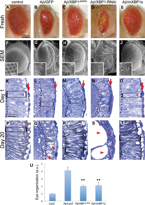
XBP1 suppresses Aβ neurotoxicity in the fly eye. Fresh eyes (A–E), scanning electron micrographs (F–J) and frontal eye sections at day 1 (K–O) and day 20 (P–T) of control flies (gmr-Gal4/UAS–GFP; A, F, K, P), flies expressing Aβ (gmr-Gal4/UAS–Aβ/UAS–GFP; B, G, L, Q) or Aβ in combination with Drosophila XBP1 (gmr-Gal4/UAS–Aβ/XBP1d08698; C, H, M, R), dXBP1–RNAi (gmr-Gal4/UAS–Aβ/UAS–XBP1–RNAi; D, I, N, S) or mXBP1s (gmr-Gal4/UAS–Aβ/UAS–mXBP1s; E, J, O, T). Compared with control flies (A and F), the eyes of flies expressing Aβ alone are small, highly disorganized and contain black, necrotic spots (B and G). The retina at day 1 is thinner (from arrow to dotted line), the photoreceptors (boxes) are very disorganized and the lenses are poorly differentiated (arrows) (K and L). By day 20, the retinas of the Aβ flies are more disorganized and vacuolated (P and Q, arrowheads). The eyes of flies co-expressing Aβ and dXBP1 (C and H) or mXBP1s (E and J) are bigger, better organized and do not contain necrotic spots. At day 1, the retinas of these flies are deeper (arrow to dotted line), the photoreceptors are better differentiated (boxes) and the lenses (arrows) are partially rescued (M and O). By day 20, these retinas maintain their organization with clearly visible photoreceptors (R and T, boxes). Flies co-expressing dXBP1–RNAi exhibit very small, disorganized and depigmented eyes (D and I) and their retinas are very thin with poorly developed lenses (N, arrow) and photoreceptors (N, box). By day 20, the retinas are mostly vacuolated (arrowheads) and no photoreceptors are visible (S). (U) Quantitation of eye phenotypes (1 = normal, 5 = small, disorganized eyes) shows that both XBP1d08698 and mXBP1s significantly reduced Aβ neurotoxicity (n= 4, P< 0.01).

