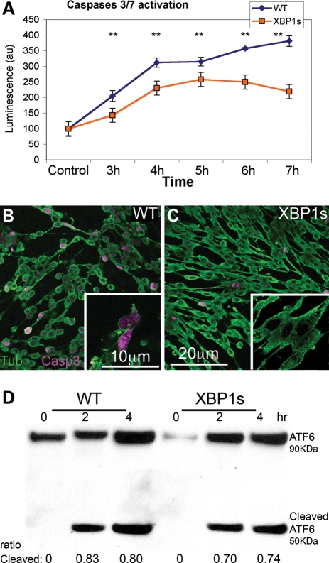
Figure 6.
XBP1s prevents Aβ-dependent caspase activation, but not ER stress. (A) Temporal analysis of caspase-3 and -7 activation in WT and mXBP1s cells treated with Aβ oligomers at 18 µg/ml. (A) WT cells (blue diamonds) progressively accumulate activated caspases over the course of the experiment. In mXBP1s cells (orange squares), activated caspase levels peak at 5 h, then decrease in the last 2h (n= 3, **P< 0.01). (B and C) Distribution of activated (act)-caspase-3 in PC12 cells treated with oligomeric Aβ at 18 µg/ml. WT cells appear small and round, and accumulate act-caspase-3 (B), whereas mXBP1s cells preserve their morphology and accumulate very little act-caspase-3 (C). (D) Western blot shows ATF6 activation in WT and mXBP1s cells treated with a subtoxic dose of Aβ oligomers (9 µg/ml). After a 4 h incubation, both WT (0.83 ± 0.0075 and 0.8 ± 0.03) and mXBP1s cells (0.7 ± 0.026 and 0.74 ± 0.023) accumulate cleaved ATF6. After a 4 h incubation, both WT and mXBP1s cells accumulate cleaved ATF6. There is no significant difference in the ratio of cleaved ATF6 between WT and mXBP1s, although mXBP1s accumulated less-activated ATF6 in two different time points.

