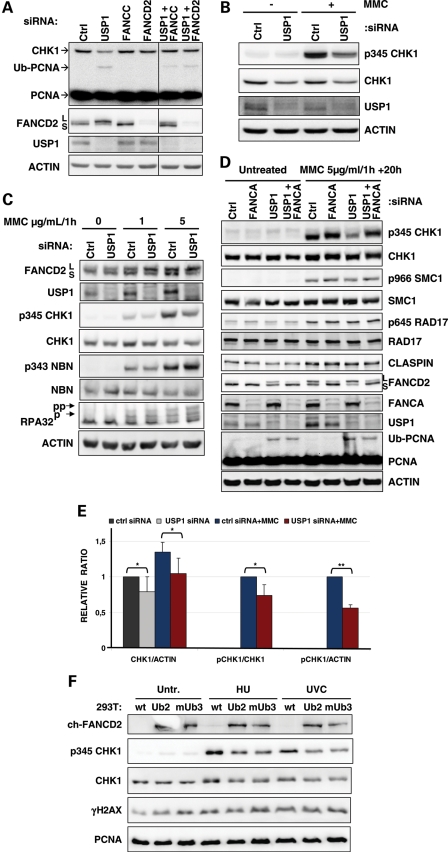
Figure 1.
USP1 depletion specifically downregulates p-CHK1 and protein levels. (A) Untreated U2OS cells were lysed 72 h after transfection with the indicated siRNAs. The lysates were analysed by western blot using the indicated antibodies. (B) U2OS cells transfected with the indicated siRNA were treated with 2 µg/ml/1h of MMC and lysed 20 h after treatment. The lysates were analysed by western blot using the indicated antibodies. (C) Two days after transfection with the indicated siRNAs, HeLa cells were treated for 1 h with the reported dose of MMC and washed before the addition of pre-warmed complete medium. Cells were lysed 20 h later, and the lysates were analysed by western blot using the indicated antibodies. (D) HeLa cells were treated with 5 µg/ml/1h of MMC 2 days after siRNA transfection, and proteins were extracted 20 h after treatment to examine the activation of several DDR proteins by immunoblotting. (E) Quantification of the ratio of CHK1 to actin, p345CHK1 to CHK1 and p345CHK1 to actin in HeLa cells treated with 5 µg/ml/1h of MMC and lysed 20 h later. Values represent the mean and SD of three (MMC treatment) to seven experiments (untreated). Student's t-test was used: *P < 0.05, **P < 0.01. (F) 293T cells stably expressing a chicken FANCD2-KR-ubiquitin fusion (clone Ub2) or FANCD2-KR-monoubiquitin fusion (clone mUb3) were treated with HU and lysed at the end of the treatment (2 mM for 30 min) or UV (5J) and lysed 1 h after irradiation, and the phosphorylation of CHK1 and H2AX was analysed by immunoblotting.

