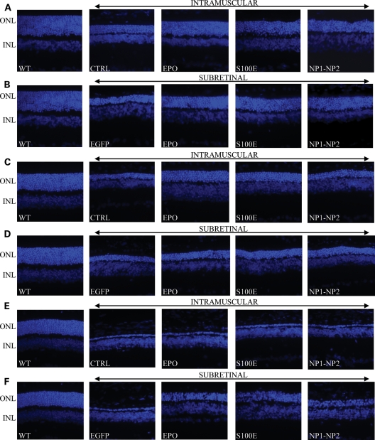
Figure 4.
Representative retinal histology in the various animal models following AAV delivery. (A, B) LD; (C, D) rds mice; (E, F) Aipl1−/− mice. LD, light damage; ONL, outer nuclear layer; INL, inner nuclear layer; WT, wild-type age-matched control animals corresponding to Lewis rats (A and B), CBA/JHsd (C and D) and C57BL/6 mice (E and F). CTRL, uninjected animals. Picture magnification is 40×.

