Fig. 4.
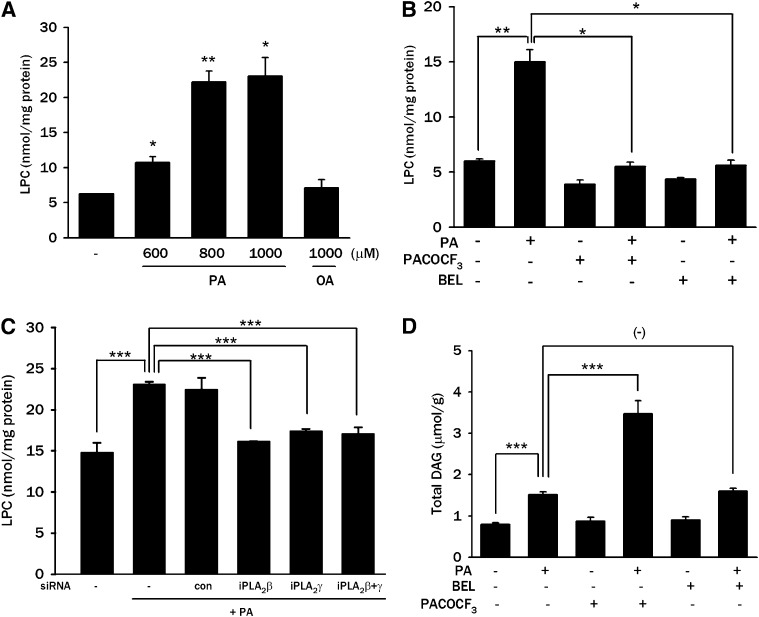
Intracellular contents of LPC and DAG after PA treatment. A: After treatment of L6 myotubes with the indicated concentrations of PA or OA for 12 h, myotubes were lysed, and intracellular LPC content was measured by an enzymatic assay as described in Materials and Methods. B: After pretreatment with 100 μM PACOCF3 or 10 μM BEL for 1 h, L6 myotubes were treated with 800 μM PA for 12 h, and intracellular LPC content was measured as in A. C: After transfection of L6 myotubes with iPLA2β siRNA, iPLA2γ siRNA, or control siRNA (Con), myotubes were treated with 800 μM PA for 12 h, and intracellular LPC content was measured as in A. D: L6 myotubes were treated with 600 μM PA for 12 h after pretreatment with 100 μM PACOCF3 or 10 μM BEL for 1 h. Content of total intracellular DAG was measured as described in Materials and Methods. The values (means ± SE) are representative of three or four independent experiments performed in triplicate showing similar tendencies. *P < 0.05; **P < 0.01; ***P < 0.005.

