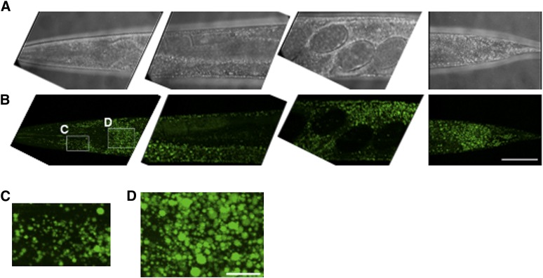
Fig. 9.
BODIPY 493/503 staining protocol stains neutral lipids in the major storage compartments, intestine and hypodermis, and in oocytes. Brightfield (A) and BODIPY 493/503 confocal images (B) of 1-day-old adult wild-type worms are shown in 3D projection of ∼25 images from a z-stack at 1 µm interval. Four segments from a wild-type worm were imaged with a 63× oil immersion objective. The anterior part is on the left. Bar, 50 µm; (C) magnified view of hypodermal lipid droplets in the head. (D) Magnified view of lipid droplets is shown in the first intestinal cells. Bar, 10 µm.

