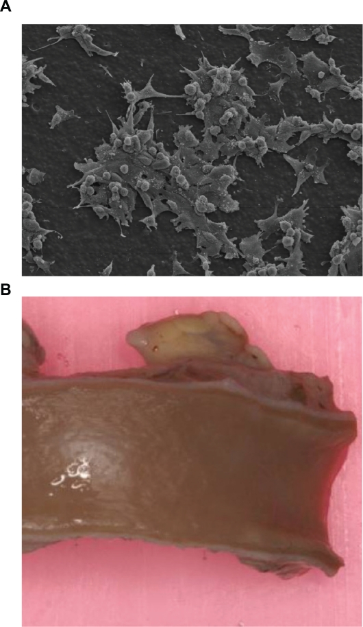
Figure 7.
A) Scanning electron microscope pictures of endothelial cell adhesion morphology on POSS-PCU showing the presence of flat, spindle-shaped cells with numerous filopodia and the absence of cell retraction. This indicates the viability and proliferation of these cells on POSS-PCU at 48 hours (320× magnification). B) An explanted POSS-PCU bypass graft demonstrated to be endothelialized and patent after a 2 year implantation in a sheep model.
Abbreviation: POSS-PCU, polyhedral oligomeric silsesquioxane-poly(carbonate-urea)urethane.

