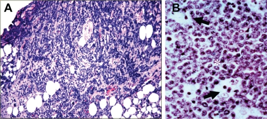
Figure 2.
A) Histopathology of the lid biopsy tissue showing inflammation and invasion of the lid tissue by large lymphocytes. B) High power magnification of the biopsy revealing the typical appearance of BL characterized by the tightly packed proliferative neoplastic lymphocytes (*) with high nuclear-to-cytoplasmic ratios interspersed with histiocytic cells (large tingible body macrophages) containing dead cell debris (arrows) yielding a “starry sky” appearance (ie, stars in a sky of B cells). (H&E; 400×).

