Figure 2.
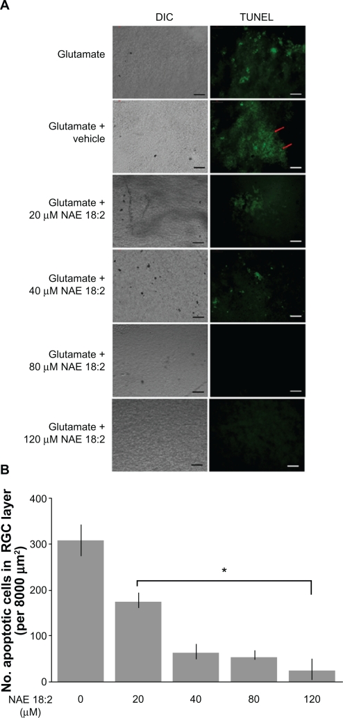
NAE 18:2 protects RGC layer neurons from glutamate excitotoxicity. A) Exposure of retinas to 100 μM glutamate resulted in a dramatic increase in RGC layer neuron death (red arrows). Preincubation of retinas with NAE 18:2 for 6 hours prior to glutamate exposure resulted in a dose-dependent decrease in the number apoptotic, TUNEL-positive RGC layer neurons, with high physiological concentrations reducing the number of apoptotic neurons. RGCs were identified by Thy 1.2 immunoreactivity and location.15,32 B) Quantitative summary data for the NAE 18:2-mediated neuroprotection from glutamate toxicity as measured by TUNEL histochemistry.
Notes: *denotes P < 0.05, as determined by a one-way analysis of variance test. Scale bar: 50 μM.
Abbreviations: DIC, differential interference contrast; NAE, N-acylethanolamide; RGC, retinal ganglion cell; TUNEL, terminal deoxynucleotidyl transferase-mediated dUTP (2′-deoxyuridine 5′-triphosphate) nick-end labeling – green fluorescence.

