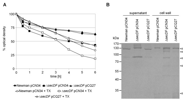
Figure 5.
Autolysis and zymogram. (A) Spontaneous and Triton X-100 (TX) induced autolysis was measured over time. (B) Autolysin zymography of protein extracts from supernatant and cell wall was performed using SDS-10% PAGE supplemented with S. aureus cell wall extract as a substrate. Dark bands show hydrolyzed cell wall and are indicated by triangles. Based on the work of Schlag et al. bands were assigned as follows in decreasing order: Pro-Atl (~130 kDa); Atl (~115 kDa); Atl-amidase (~84 kDa) or part of the propeptide (62-65 kDa); Sle1/Aaa (~33 kDa) [35].

