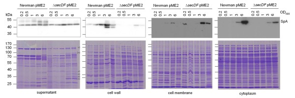
Figure 7.
Subcellular localization of SpA. Expression and localization of SpA was monitored in the Newman pME2 background during growth. Upper panels show Western blots of SpA. Longer exposure times were required for detection of SpA in cell membrane and cytoplasm. Bottom panels show Coomassie stained gels. Bands of stronger expression in the mutant are indicated by triangles.

