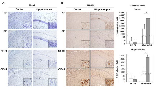
Figure 2.
Neuropathological examination. (A) Nissl staining showed no difference between the NF pups and OF pups in the cortex and hippocampus on P7, but 24 hours post-HI, the OF-HI pups had more neuronal damage than the NF-HI pups. (B) TUNEL staining revealed that the OF-HI pups had significantly more TUNEL-(+) cells in the cortex and hippocampus than the NF-HI pups. n = 4. *p <0.05. Scale bar = 100 μm (A, B), and = 10 μm in the insets (A, B).

