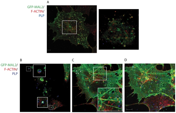
Figure 7. Colocalization of PLP and GFP-MAL2 with actin cytoskeleton.
GFP-MAL2/HOG cells cultured in DM at 37°C for 2 days were fixed and processed for confocal triple-labelled indirect confocal immunofluorescence analysis with anti-PLP monoclonal antibody and Phalloidin-TRITC to stain F-actin. Monoclonal anti-PLP antibody was detected using an Alexa Fluor 647 secondary antibody. A. Most of GFP-MAL2-positive vesicles contain F-actin. The large image is a projection of confocal planes. The small image is an enlarged 0.8 µm confocal plane corresponding to the square. B. Colocalization of PLP, GFP-MAL2 and F-actin can be observed in some vesicles (squares). C. GFP-MAL2-positive tubular reticulum colocalizes with F-actin (squares) and PLP (arrows). Structures showing colocalization of GFP-MAL2 with F-actin appear yellow, and GFP-MAL2 with PLP appears cyan. B and C are 0.8 µm confocal planes from the same image, and D is the projection of all confocal planes corresponding to that image. Scale bar = 5 µm.

