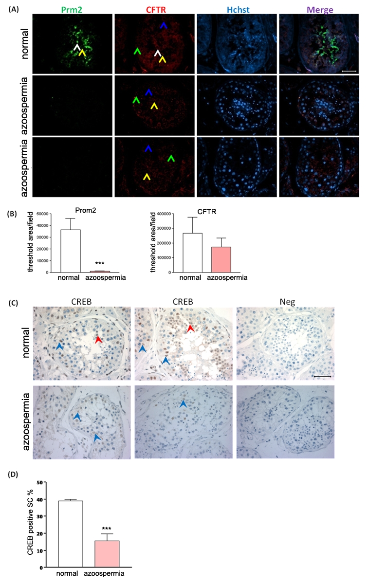
Figure 4. Reduced CFTR and CREB expression in human azoospermia testes.

(A) Representative immunoinfluoresence staining of CFTR (red) and Protamine-2 (green) in normal human (n = 5) and azoospermia patients (n = 6) testes, counterstained with Hoechst (blue). In the normal human testes, Protamine-2 positive signals are localized in the round spermatids (RS, yellow arow) and elongated spermatids (ES, white arrow) (Stages I–II); CFTR positive signals are localized in pachytene spermatocytes (P, blue arrow), RS and Sertoli cells (SC, green arrow) (stages I–VI), especially in RS. In azoospermia testes, no positive signal of Protamine-2 and weak signals of CFTR are detected (Magnification: 400x). Bar = 50 µm. (B) Statistic analysis of protamine-2 and CFTR positive signal area in normal (n = 5) and azoospermia (n = 6) human testes samples (***:p<0.001). (C) Immunohistochemistry of CREB in normal human and azoospermia testes. In normal human testes, CREB positive signals are localized in RS (red arrow) and SC (blue arrow). CREB inmmunoreativity in SC is decreased in the testes with spermatogenic arrest obtained from azoospermia patients. Bar = 50 µm. (D) Statistic analysis of CREB positive Sertoli cell percentage in normal (n = 5) and azoospermia (n = 6) human testes (***:p<0.001). Statistic data are shown as mean±SEM.
