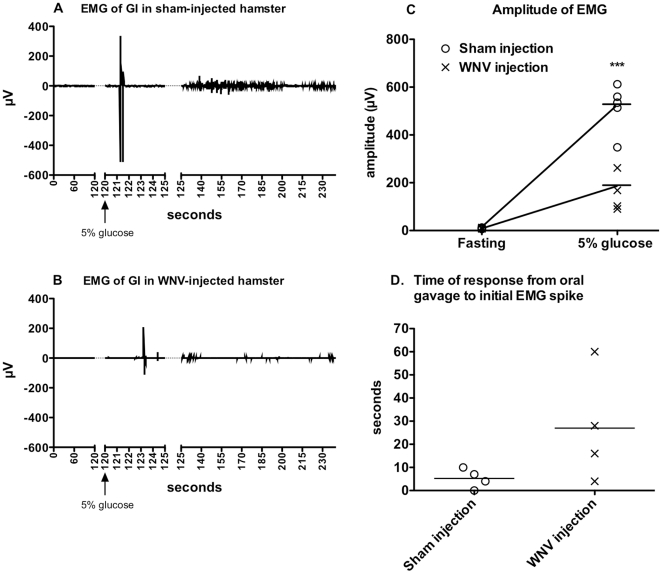
Figure 2. Electromyography (EMG) of intestines in WNV-infected hamsters.
Hamsters were surgically implanted with telemetry transmitters with electrodes attached to the duodenum just below the pyloric sphincter, and then injected s.c with WNV or sham. At 9 days after viral challenge, EMGs were measured continuously for 4 min in isoflurane-anesthetized hamsters that had fasted for 15 hr. At 2 min (arrows), hamsters were orally gavaged with 1.5 mL of 5% glucose. Representative EMG tracings of A) sham-injected and B) WNV-injected hamsters. C) Mean amplitudes ± SD from 0–2 min. (fasting) and 2–4 min. (***P≤0.001 using a two-way t test.)

