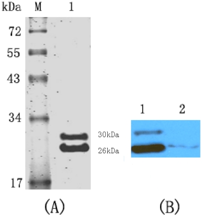
Figure 2. SDS-PAGE and western blotting analysis of the protein of purified Fab094.
(A) Proteins were separated by 12% SDS-PAGE and stained with Coomassie Brilliant Blue. Lane 1 shows the protein of purified Fab094. Lane M contained the molecular-mass markers. (B) Western blotting analysis of Fab094 with HRP-conjugated anti-human IgG (Fab-specific). Lane 1 shows the ultrasonic supernatant of Fab094 with HRP-conjugated anti-human IgG; lane 2 was the culture supernatant of Fab094 with HRP-conjugated anti-human IgG.

